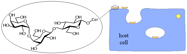
The Shiga-like toxins recognise target cells by binding to the carbohydrate portions of certain glycolipids on the cell surface. Glycolipids are membrane components containing a fatty acid portion, which is buried inside the membrane, and an exposed carbohydrate portion. Most members of the Shiga-like toxin family bind to a particular glycolipid called Gb3; its carbohydrate component consists of the three linked sugar groups shown below. (Pig edema toxin is an exception. It binds to Gb4, which has an extra sugar group at the end of the carbohydrate chain.) In the picture of Gb3, Cer indicates the ceramide portion that is buried in the interior of the membrane.
The damage caused by these toxins is determined partly by which cells are exposed to toxin, but also largely by the content of Gb3 in the cell membranes. If the toxin does not enter the bloodstream, damage is limited to the digestive tract, particularly the ascending colon. Damage to the kidneys, when the toxin does enter the bloodstream, can be explained by the high content of Gb3 in the affected cells in the glomeruli of the kidneys.
Carbohydrate binding is the first step in toxin action, which makes it a good point to intervene with drugs. In addition, it occurs outside the cell, which means that drugs interfering with binding wouldn't have to get inside cells, a serious hurdle for potential drugs. To explore the possibility of designing drugs that interfere with cell binding, we looked at binding of the trisaccharide from Gb3.
|
Back Structure of SLT-I B-pentamer |
Up Back to top |
Next Sugar binding to SLT-I |