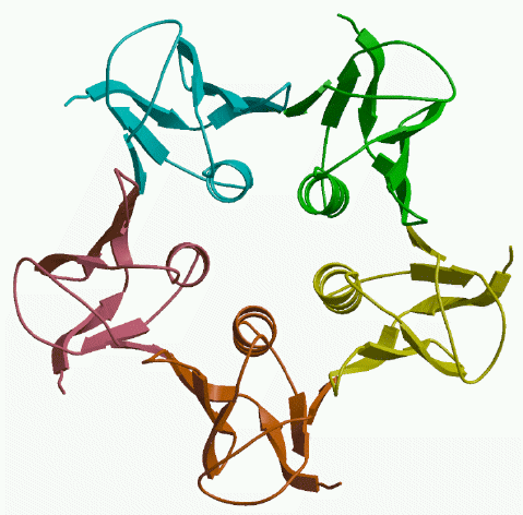
We began our study of this toxin by looking at the 3D structure of the B-subunit on its own. Five copies of the B-subunit protein associate to form a pentamer with five-fold symmetry, as shown in the figure below. This structure turns out to resemble that of the B-subunit of cholera toxin, which was a bit of a surprise because the amino acid sequences are very different. But this means that we can draw some lessons from what has been learned about the binding of cholera toxin to its receptors.
This structure, and the other structures discussed on these web pages, were determined using the technique of protein X-ray crystallography. If you want to understand how this works, you could take a look at notes from our lecture series on protein crystallography. The image in the picture above is a schematic drawing of the polypeptide chain of the toxin, with beta-strands shown as broad arrows and alpha-helices shown as coiled ribbons.
|
Back AB toxins |
Up Back to top |
Next Receptors for Shiga-like toxins |